There was once a patient experiencing a serious complication during a brain blood vessel surgery due to the lack of detailed anatomic visualization. This specific case shows the importance of technology in assisting surgeons to understand complex structures of brains before conducting any form of surgery.
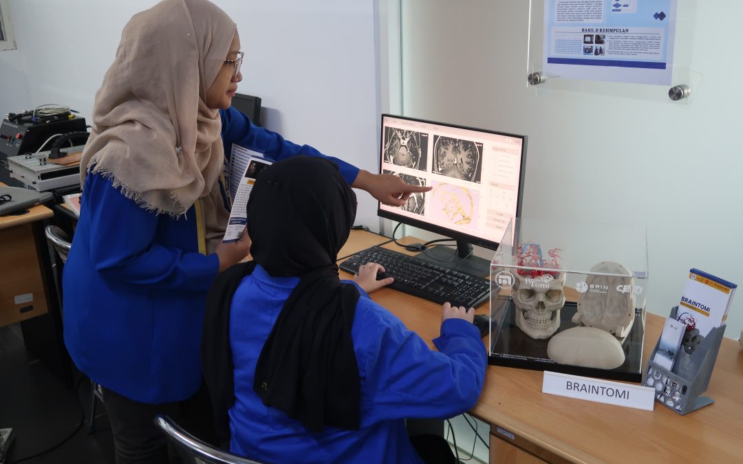
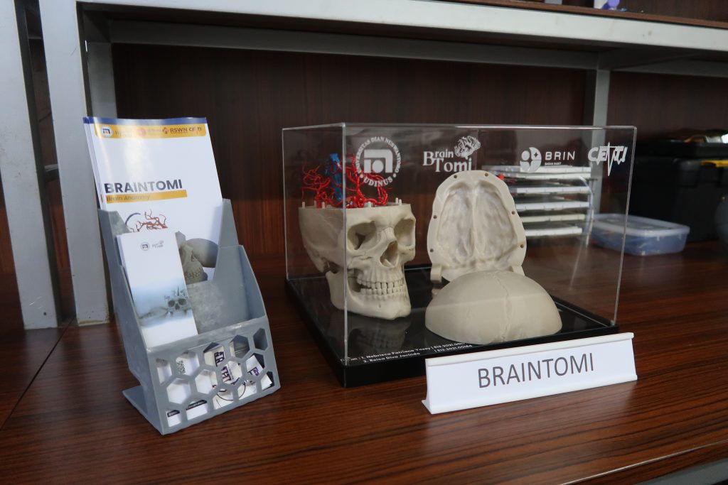
In response to this problem, a faculty member from the Biomedical Engineering Department at Universitas Dian Nuswantoro (Udinus), Menik Dwi Kurniatie, S.Si., M.Biotech., developed a three-dimensional (3D) model of blood vessels in the human brain. The model was named Brain Anatomy or ‘Braintomi’ for short. Its primary purpose is to improve surgeons’ competence in planning so that surgeries can be done more accurately.
The research has been published in the Journal of Electronics, Electromedical Engineering, and Medical Informatics in the January 2025 edition. During the research, Menik and her team utilized Magnetic Resonance Imaging technology, which was combined with 3D printing technology.
“The model is designed so surgeons can understand more accurately the structure of blood vessels. It also serves as a way for surgeons to conduct a simulated surgery before doing the real one. We have seen that 2D-based Magnetic Resonance Imaging (MRI) does not provide a clear enough spatial description, especially for complex cases. For that reason, we designed this model to be three-dimensional instead,” she uttered.
The research process involved an image segmentation from the MRI data. The data would then be processed using a particular software to form a digital model depicting blood vessels in the brain. The model would then be printed using technology known as Stereolithography Apparatus (SLA) and Fused Deposition Modeling (FDM).
“As a result, we could fully replicate the blood vessels in such detail that they look like real blood vessels. This technology is expected to reduce the risks during surgery. By identifying challenges that might happen, we could essentially determine the best strategies to face them,” Menik uttered.
As additional information, the innovation also involved two Biomedical Engineering students from Udinus, serving as their final projects. Those students were Nebrisca Patriana Yossy and Reica Diva Jacinda.
During its creation, Braintomi also received support from various parties, including the National Research Innovation Agency, the KRMT Wongsonegoro Hospital, and the Center for Medical Technology Innovation (Cemti).
Improving the Quality of Surgeons
During her research, Menik also involved her fellow faculty members, one of which was a lecturer from the Medicine Faculty at Udinus, dr. Andreas Wilson Setiawan, M.Kes., as the validator of blood vessel anatomy. According to dr. Wilson, the research could be adopted in various medical institutions to improve surgeons’ competence, as well as medical students.
He emphasized that the innovation could open a bright opportunity for the medical field to improve the accuracy and surgery operational procedures in the future. “Hopefully, soon enough, this model could be further developed with materials that resemble human brain network, allowing it to provide a more realistic simulation for surgeons,” dr. Wilson concluded. (Humas Udinus/Haris. Foto: Humas Udinus)